Why I-MED Radiology
At I-MED Radiology, when you need answers, we are here to provide the highest quality care and deliver the most accurate diagnostic outcomes ...

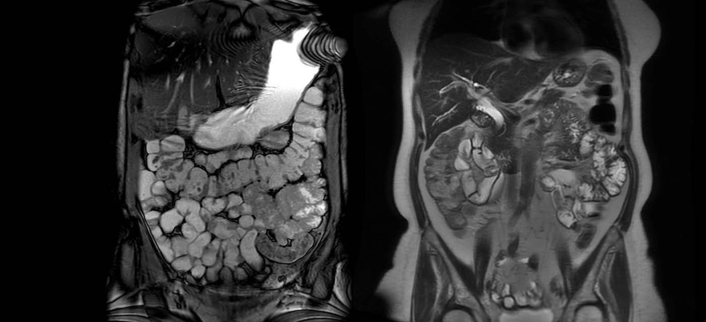
The procedure involves filling the bowel with fluid that will show up brightly on the images and makes the small bowel stand out. This study typically involves the injection of a special dye (often called ‘contrast medium’ or ‘contrast agent’) into the veins during the scan. The contrast highlights the bowel wall on the images. This contrast medium is known as gadolinium.
Patient preparation can vary but usually, you will asked to fast or have only a light breakfast prior to the procedure. Blood tests may be requested to see if you have any kidney problems.
Radiologists typically use an injection to slow down the normal movements/contractions of your bowel (as these contractions can blur the MRI pictures). This injection will usually be called Buscopan (hyoscine) or glucagon. You may also need to have a gadolinium injection, which is injected via a cannula placed prior to the procedure.
The fluid you drink for enterography may make you feel slightly nauseous. Occasionally the fluid may also cause mild diarrhea after the test.
Your doctor will receive a written report on your test as soon as is practicable.
It is very important that you discuss the results with the doctor who referred you so they can explain what the results mean for you.
Most results are normal. Occasionally, small changes are seen that need further review.
If your results are normal you will be able to return for routine screening (usually every 2 years). If your results are uncertain or show changes you may need to consider additional imaging (ultrasound, or biopsy) in discussion with your referring doctor.

This information has been reviewed and approved by Dr Ronald Shnier (I-MED Chief Medical Officer).
This information has been reviewed and approved by Dr Ronald Shnier (I-MED Chief Medical Officer).